Our study will take place in a lab where the fungus P. destructans will be grown in petri dishes on sabouraud dextrose agar, which supports the growth of fungi (Thermo Fischer Scientific, 2020). There will be five treatments tested to inhibit growth of P. destructans: a strain of Pseudomonas spp isolated from E. fuscus (PF1) (biological), a strain of Pseudomonas spp isolated from M. lucifugus (PF2) (biological), chitosan (biological), B23 (chemical), and PEG 8000 (chemical). Each treatment will be grown on its own petri dish to prevent cross contamination, with a 5mm plug of the fungus placed in the center of each dish (Figure 3). The cultures will be grown for 13 days, aligning with McDonald et al’s (2019) methods, under three different conditions to simulate the temperature and humidity of different bat hibernacula: 5°C, 10°C, and 2°C (also called sites T5, T10, and T2); this is based on Blehart et al. (2009) and Grieneisen (2011)’s information on the optimal temperature growth range of P. destructans. Humidity will be kept constant at 95% as bats tend to find areas of high humidity (ranging from 90-100% humidity) for hibernation (Grieneisen, 2011). There will be five repetitions of each treatment done at each site for a total of 75 samples. After the 13-day growth period, the samples will be assessed using a 50 square grid centered on each of the fungus plugs to determine the percentage of inhibition relative to the control (Figure 4). The samples will then be checked again 10 days later, using the same method as above to measure inhibition, to see if any regrowth occurred.
Post-data collection, statistical analyses were performed in R to compare the effectiveness of the five treatments against P. destructans (R Core Team, 2019). To determine this, five packages were installed in R: Multcomp (Hothorn et al., 2008), multcompView (Graves et al., 2019), Sciplot (Morales et al., 2020), Plyr (Wickham, 2011), and Emmeans (Russell, 2020), and a multi-factor ANOVA and effect size statistics were conducted (R Core Team, 2019).
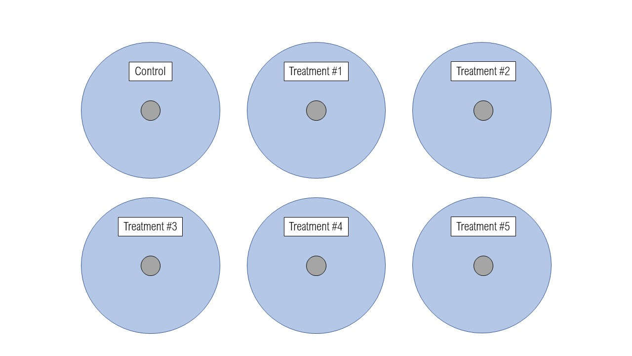
Figure 3. A diagram of the petri dishes in which each treatment will be applied for testing, with the grey circle representing a 5 mm fungus plug.
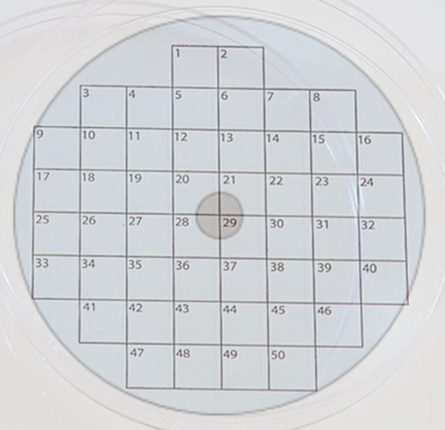
Figure 4. A petri dish from Figure 3 with a sticker grid added for counting. (Roth, n.d.). Retrieved from: https://tinyurl.com/yycasv5h
